Anamnese:
Eine 72-jährige Patientin klagte über starke Schmerzen in der rechten Hüfte beim Gehen. Ein Fortbewegen war nur noch mit Gehhilfe möglich. Bei der Bewegung des rechten Beines unter normaler Belastung spürte die Patientin eine Art "Schnappen" des Gelenks.
Die Patienten weist bereits eine längere Krankheitsgeschichte mit mehreren operativen Eingriffen der betroffenen Hüfte auf, darunter eine Fraktur des Oberschenkelhalses rechts (1991), ein totaler Hüftgelenksersatz mit Pfannen- und Schaftprothese (1996) und ein Wechsel der Gelenkkugel mit gleichzeitiger Wiederherstellung der natürlichen Beinlänge (2015).
Untersuchung und Diagnose:
Die radiologische Untersuchung zeigte eine deutlich außermittige Stellung der Schaftprothese zur Pfannenprothese. Auffällig waren außerdem die weit in das Weichteilgewebe des Beckens hineinragenden Schrauben zur Fixierung der Pfannenprothese. Die Orientierung und Lage der Pfannenprothese wurde als nicht optimal bewertet. Eine zeitlich bedingte Lockerung beider Prothesen konnte nicht ausgeschlossen werden.
Therapie:
Aufgrund des Alters der Patientin, des Alters der Prothesen und der ungünstigen Orientierung der Hüftpfanne fiel die Entscheidung für einen Austausch der Hüftpfannenprothese.
Ergebnis:
Aufgrund von Lage und Alter der bestehenden Pfannenprothese sowie der beiden in das Becken ragenden Schrauben wurde ein 3D-Modell erstellt, welches die Problematik verdeutlichte. Mit Hilfe des Modells konnte die Situation optimal eingeschätzt und präoperativ vorbereitet werden.
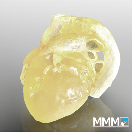
Digitale Rekonstruktion (Virtual 3D model)
Hip-TEP revision
Case history and admission findings:
A 72-year-old patient suffered severe pain in the right pelvis when walking. She can only move by using a walking aid. During a movement of the right leg under normal load, the patient felt a “snapping” of the hip joint.
The patient already has a long medical history with several surgical procedures in the hip area, including a fracture of the femoral neck on the right side (1991), a total hip replacement with acetabular and shaft prosthesis (1996) and a change of the ball joint with simultaneous adjustment of the natural leg length (2015).
Investigations and diagnosis:
The radiological investigation revealed an eccentric position of the shaft prosthesis to the acetabular prosthesis. The screws for the fixation of the acetabular prosthesis extended too far into the soft tissue of the pelvis. The orientation and position of the acetabular prosthesis was rated as not optimal. A loosening of both prostheses could not be excluded.
Treatment:
Due to the age of the patient, the age of the prosthesis and the unfavorable position of the acetabulum, the treating physician decided for an exchange of the acetabular prosthesis.
Result:
Due to the position and age of the acetabular prosthesis as well as the screws the treating physician was provided with a 1:1 scale 3D printed model of the pelvis. This model helped the surgical team to analyze the complexity. With the aid of the model the procedure could be prepared preoperatively.
Patientendaten
Alter: 72 Jahre
Geschlecht: weiblich
Diagnostik:
- CT
- Röntgen
Patient data
72-year-old female
Diagnosis:
- CT scan
- X-ray
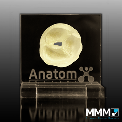
Präoperative Planungsmodelle
(Printed 3D model)